Veterinary Microscope with 100X DRY Objective - NEVER USE OIL AGAIN
What is Dark Field Microscopy?
When almost anyone is first introduced to microscopes, the instrument they are shown is the traditional, familiar bright field microscope. The term ‘bright field’ refers to the way objects to be viewed are illuminated. In a bright field microscope, light is passed from a point below or beneath the specimen, through the specimen, on up through the objective lens and eventually to the instrument’s eyepiece. What one ordinarily sees through the eyepiece of a bright field microscope is a brightly-lit circle (or field), with the specimen somewhere within that circular field of view.
As an example, viewing a thin slice of a plant stem, prepared, stained and mounted on a glass slide and placed on the viewing stage of such a microscope, might reveal an orderly structure of cells – darker lines, granules and so on, against a bright field.

This system works very well for quite a number of applications. In some cases, though, it doesn’t allow the viewer to see what he or she really needs to see.
For example, if one needed to be able to view cells or microorganisms that had not been stained, one would be able to see little or no detail, using bright field illumination. Since the objects under study are transparent (or very nearly so) you might see nothing more than vague outlines.

One solution to this type of viewing requirement is the Dark Field Microscope. The microscope itself needn’t really be any different than one used in bright field viewing. The difference is in how specimens are illuminated. In a dark field system, objects in the field of view are lit from the sides, by what amounts to a hollow cone of illumination, projecting upward and outward from below the stage. As described earlier, in a bright field lighting arrangement, light passes through the specimen – usually from below. In dark field illumination, light coming from directly below the viewing stage is blocked and/or redirected so that the only light falling on the field of view comes from all sides. What the viewer sees with this type of lighting is brightly lit objects (lit obliquely, from all around) against a dark background.

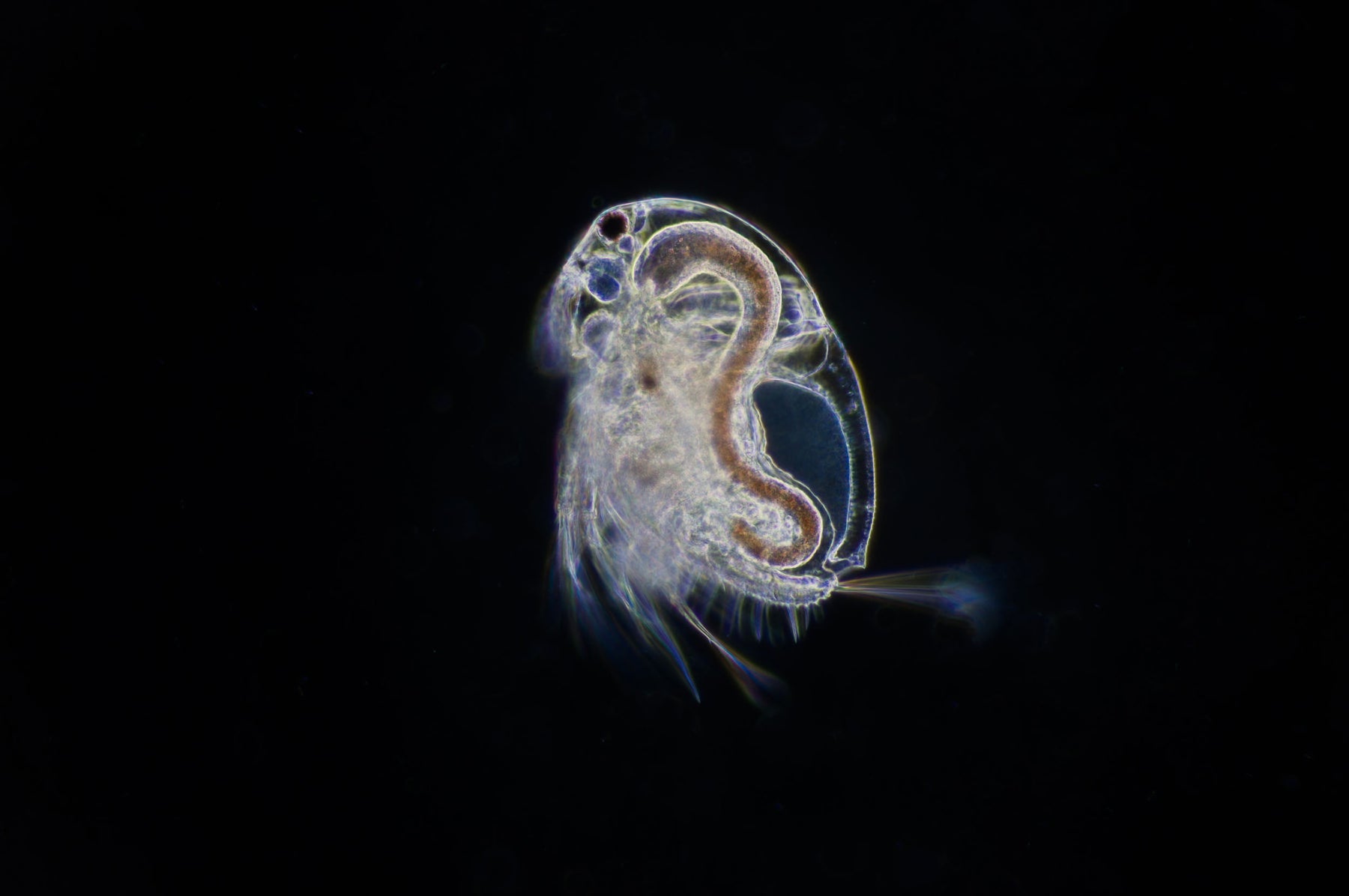
With this lighting method one cannot ordinarily see much detail of internal structures – but external textures, features and structures stand out strongly. It also allows excellent viewing of minute organisms – tiny insects or sea life, for example, or single-celled microorganisms – even while they are still living.

Red & white blood cells under the Optico BM2000 Dark Field Microscope
Dark field microscopes are also ideally suited for an emerging technology and practise in the field of health care, known as live blood analysis. In live blood microscopy, a fresh, still-living sample of blood is viewed using a dark field microscope; the blood’s various living components can be seen and assessed, along with any foreign objects or debris that might be present. Even the state of the fluid itself can be observed.

As the use of dark field microscopes has expanded into more and more areas of application, a variety of methods and devices (known as condensers) for providing dark field illumination have also been developed. Each has its own configuration of lenses and reflective surfaces to direct an inverted cone of light upward to the viewing stage, at varying intensities and angles.

Returning to the field of live blood analysis, perhaps the most effective such device for use with a live blood microscope is the combination of using an OIL Darkfield Condenser, with a 100x Oil Immersion Darkfield Objective & Iris Diaphragm, 3W Koehler Illumination System & Professional Quality 1080P HD Digital camera as shown below:

Please feel free to contact us if you require any further assistance in choosing the right microscope for your requirements.